

The University of Oxford offers an extensive catalogue of equipment for the essential tools of Structural Biology; cutting-edge mass spectrometry, microscopy and biophysical analysis, with characterisation of high containment level pathogens.
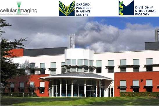
Oxford Particle Imaging Centre (OPIC) is part of the Division of Structural Biology (Strubi). OPIC houses state-of-the art equipment for cryogenic electron microscopy (cryo-EM/cryo-TEM), including Titan Krios and a Glacios instruments, both fitted with direct electron detectors, with access to auxiliary equipment for sample preparation. Central to OPIC are biosafety containment laboratories which facilitate the study of infectious viruses relevant to human and animal health, and it is possible to perform HG3 category pathogen study using our Titan Krios microscope.
Cellular Imaging Core Facility (CICF) is a major hub for light microscopy providing imaging solutions from the organismal to the molecular level, offering valuable insight ahead of and complementary to structural work. We provide live cell and high-content imaging modalities allowing users to better understand proteins in context of live cells, thus providing the benefit of temporal resolution. Ongoing support and guidance is provided at all stages of light microscopy from experimental design through to image analysis.
Oxford Mass Spectrometry Centre offer a wide range of services and several pieces of equipment to perform native mass spectrometry as well as hydrogen-deuterium exchange mass spectrometry. As no two experiments are the same, we offer services tailored to your needs. OMSC also now offer mass photometry, a relatively new method for analysing molecules. It enables the accurate mass measurement of single molecules in solution, in their native state and without the need for labels. This approach opens up new possibilities for bioanalytics and research in the functions of biomolecules.
The Molecular Biophysics Suite provides a wide range of biophysical techniques for researchers and commercial users. The facility specialises in the characterising recombinant proteins and their interactions in solution and is therefore well equipped with instrumentation to characterise the solution state, interactions and kinetics.
The Cellular Imaging Core Facility (CICF) is a major hub for microscopy in Oxford, providing imaging solutions from the organismal to the molecular level. The CICF is much more than a point of use facility. In addition to excellent quality tailored user training and ongoing support on all systems covering a wide range imaging modalities, we also offer advice and guidance throughout each stage of imaging projects. We strongly encourage users come to the core at the project conceptualisation stage, we then work with them through planning, sample preparation, imaging, data analysis, figure preparation and finally publication.
The Oxford Particle Imaging Centre (OPIC) provides state-of-the-art cryogenic electron microscopy for single-particle analysis, cryo-electron tomography (cryo-ET), cryo-correlative light and electron microscopy (cryo-CLEM), and in situ structural biology. Our workflows support specimens ranging from purified macromolecular complexes and viruses to intact cells, organoids and tissues. OPIC is available to researchers across the University of Oxford and to external academic and industrial users.
OPIC provides comprehensive infrastructure for cryo-EM sample preparation and optimisation, including Vitrobot and Leica GP2 plunge-freezing devices, high-pressure freezing, and PRIMO (Alvéole) micropatterning for controlling cell growth and positioning on EM grids.
For cryo-EM screening and data collection, OPIC houses a 200 kV Thermo Fisher Scientific Glacios cryo-TEM for sample screening, grid optimisation and initial data collection, and a 300 kV Titan Krios G3i cryo-TEM, equipped with a Falcon 4i direct electron detector and Selectris-X energy filter, for high-resolution single-particle and cryo-ET data acquisition.
For in situ structural biology, OPIC operates two complementary cryogenic FIB-SEM platforms. The Thermo Fisher Scientific Arctis Cryo-Plasma FIB enables automated, high-throughput preparation of cryo-lamellae from vitrified cells. Its integrated fluorescence light microscope (iFLM) supports fluorescence-guided target selection and verification during milling, while Autoloader-based sample handling enables streamlined transfer between cryo-FIB preparation and cryo-TEM data collection. The Thermo Fisher Scientific Aquilos 2 FIB-SEM, also equipped with integrated fluorescence imaging, supports conventional cellular lamella preparation as well as cryogenic lift-out, extending in situ cryo-ET to thicker and more complex specimens, including organoids and tissues.
Together, these technologies provide integrated workflows from sample preparation and fluorescence-guided targeting through cryo-FIB milling to high-resolution cryo-ET, alongside conventional single-particle cryo-EM workflows from initial grid optimisation to high-resolution data collection.
A distinctive capability of OPIC is that its instrumentation is embedded within ACDP Category 3 and DEFRA Category 4 containment laboratories, enabling cryo-EM studies of approved infectious pathogens relevant to human and animal health. OPIC therefore brings together single-particle cryo-EM, cellular cryo-ET, cryo-CLEM and high-containment structural biology within a single facility.
HDX Mass Spectrometry gives information of the exchange of hydrogen for deuterium on the protein backbone. Uptake rate is determined by each site’s solvent accessibility. Comparing the extent of deuterium exchange in a ‘apo’-protein with a target (agonist, antagonist, inhibitor, lipid etc) bound protein can provide information about conformational changes, active sites and important binding residues.
Instruct provides full use of the Strubi-crystallisation facility for setting up crystallisation experiments and monitoring these experiments via a web-based graphical user interface (GUI). Facilities include setting up screening experiments and also various optimisation methods including grid screens and additive screens. Experiments are typically performed in 100nL sample + 100nL reagent vapour-diffusion format.
Mass Photometry is an interferometric microscopy technique which allows single molecule mass measurement of biological molecules (proteins, DNA, antibodies, etc) by detecting minute changes in the reflected light caused by changes in dielectric constant of the medium as the molecule approaches and binds to a surface. The method has myriad applications to biophysical systems, ranging from rapid in lab mass measurements to tracking single molecules moving across a substrate.
Native Mass Spectrometry is the utilisation of soft ionisation methods and biological like sample conditions to allow a mass spectrum of a natively folded (i.e. not denatured) protein to be collected. Coupled with high resolution and highly sensitive detectors, this allows for: accurate mass, stoichiometry and, in some cases, small molecule/lipid binding partner determination.